Fluigent introduces Aria our automated solution for cellular perfusion or timed injection protocols. Aria allows…
Customer paper on nuclear deformations
A microfluidic device for characterizing nuclear deformations
By Andrew C. Hodgson, Christophe M. Verstreken, Cynthia L. Fisher, Ulrich F. Keyser, Stefano Pagliara and Kevin J. Chalut from the University of Cambridge and University of Exeter, UK.
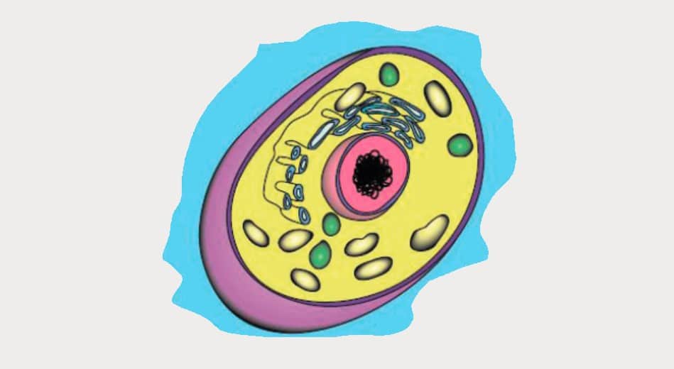
Cell nuclei experience and respond to a wide range of forces, both in vivo and in vitro. In order to characterize the nuclear response to physical stress, we developed a microfluidic chip and used it to apply mechanical stress to live cells and measure their nuclear deformability. The device design is optimized for the detection of both nucleus and cytoplasm, which can then be conveniently quantified using a custom-written Matlab program. We measured nuclear sizes and strains of embryonic stem cells, for which we observed negative Poisson ratios in the nuclei. In addition, we were able to detect changes in the nuclear response after treatment with actin depolymerizing and chromatin decondensing agents. Finally, we showed that the device can be used for biologically relevant high-resolution confocal imaging of cells under compression. Thus, the device presented here allows for accurate physical phenotyping at high throughput and has the potential to be applied to a range of cell types.
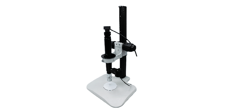
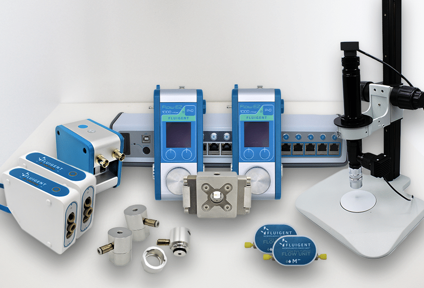
This work was made possible thanks to our MFCS™-EZ, used with MAESFLO™ to inject cell suspensions in incubating conditions (37°C and 7% CO2)
For more information: doi: 10.1039/C6LC01308B or Lab Chip, 2017, 17, 805-813.
Related Posts