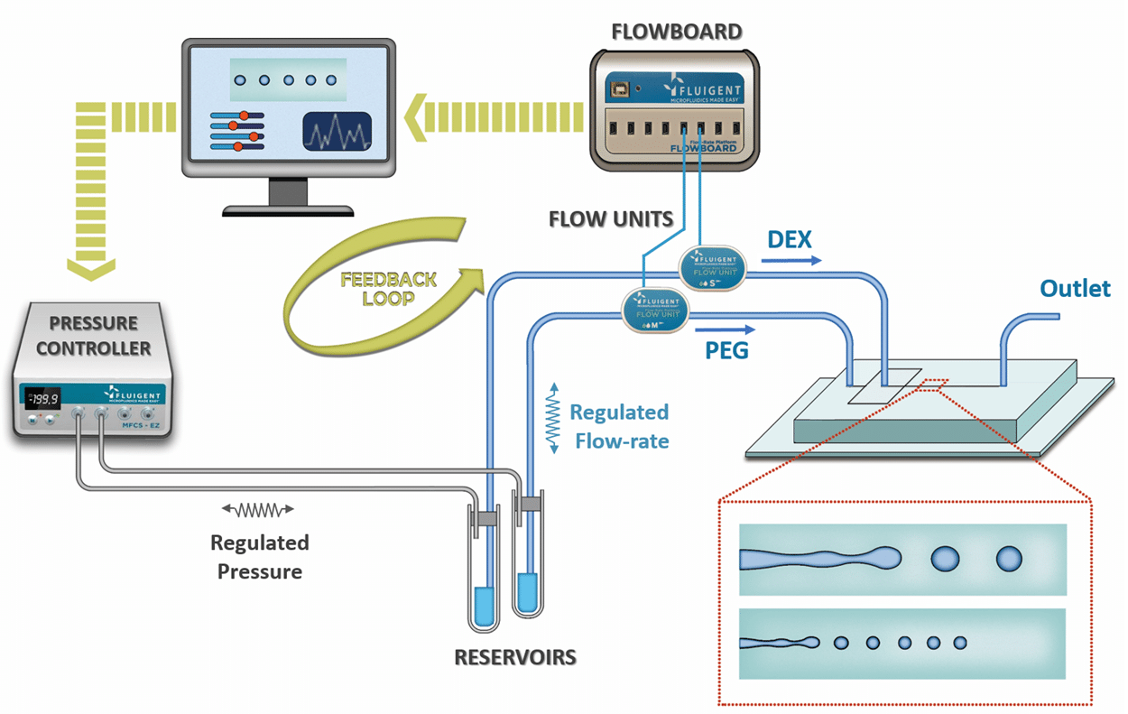
Microbead-based microfluidics (droplet-based microfluidic using specific gel as dispersed phase) is a powerful technic which…

Customer paper on mechanophenotyping cells
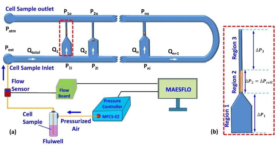
A combined experimental and theoretical approach towards mechanophenotyping of biological cells using a constricted microchannel
By
We report a combined experimental and theoretical technique that enables the characterization of various mechanical properties of biological cells. The cells were infused into a microfluidic device that comprises multiple parallel micro-constrictions to eliminate device clogging and facilitate characterization of cells of different sizes and types on a single device. The extension ratio ? and transit velocity Uc of the cells were measured using high-speed and high-resolution imaging which were then used in a theoretical model to predict the Young’s modulus Ec = f(?, Uc) of the cells. The predicted Young’s modulus Ec values for three different cell lines (182 ± 34.74 Pa for MDA MB 231, 360 ± 75 Pa for MCF 10A and, 763 ± 93 Pa for HeLa) compare well with those reported in the literature from micropipette measurements and atomic force microscopy measurement within 10% and 15%, respectively. Also, the Young’s modulus of MDA-MB-231 cells treated with 50 ?M 4-hyrdroxyacetophenone (for localization of myosin II) for 30 min was found out to be 260 ± 52 Pa. The entry time te of cells into the micro-constrictions was predicted using the model and validated using experimentally measured data. The entry and transit behaviors of cells in the micro-constriction including cell deformation (extension ratio ?) and velocity Uc were experimentally measured and used to predict various cell properties such as the Young’s modulus, cytoplasmic viscosity and induced hydrodynamic resistance of different types of cells. The proposed combined experimental and theoretical approach leads to a new paradigm for mechanophenotyping of biological cells.
This work made possible thanks to our MFCS™-EZ with MAESFLO™ and our FRP, to inject the cell solutions into their system.
For more information: doi: 10.1039/C7LC00599G or Lab Chip, 2017,17, 3704-3716.


